
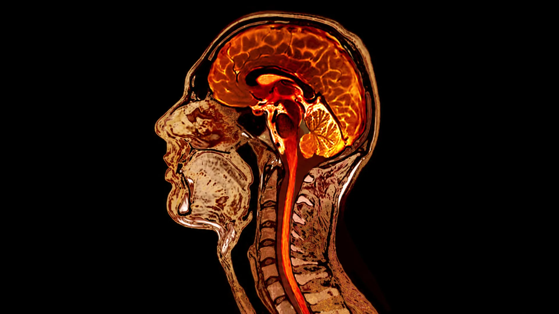




Both CT and MRI scans provide a detailed view of the inside of your body, but they’re not the same technology, nor do they create images in the same way.
If you’ve ever had a physician recommend advanced imaging, you may have wondered why they ordered a CT scan instead of an MRI, or vice versa. While both tests allow us to see structures inside the body that we can’t evaluate through an external physical exam or blood work alone, they excel at showing different structures and answering different clinical questions.
A CT scan uses ionizing X-rays to rapidly create detailed cross-sectional images of your body, which can help evaluate your lungs, bones, blood vessels, internal bleeding, and more. MRI, on the other hand, uses powerful magnetic fields and radiofrequency energy rather than ionizing radiation and is particularly valuable for visualizing many soft tissue details. This can be especially useful if a physician suspects something like a herniated disc, as an example. While CT may adequately visualize a larger herniated disc, MRI provides better soft tissue detail and contrast allowing better visualization of small herniated discs, individual nerve roots and the spinal cord itself.
That doesn’t make MRI automatically “better” or CT inherently riskier. CT may provide information that MRI can’t provide as effectively for a particular clinical question, and the reverse can also be true.
What is the difference between a CT scan and an MRI?
A CT, or computed tomography, scan takes a series of X-ray images from different angles and uses computer processing to create detailed cross-sectional images. It’s fast, noninvasive, and especially helpful when physicians need to quickly evaluate structures such as the lungs, bones, blood vessels, and other internal organs. [1]
MRI, or magnetic resonance imaging, creates images using a strong magnetic field, radiofrequency energy, and computer processing. It doesn’t use X-rays or ionizing radiation. MRI can provide excellent contrast between different types of soft tissue, making it valuable for getting detailed views of the brain, spinal cord, joints, muscles, and many internal organs. [2]
There are a few practical differences, too. CT scans are generally faster. MRI examinations often take longer and require you to remain still inside the scanner, which can be challenging for people with claustrophobia. The powerful MRI magnet also means patients must be carefully screened for certain implants, medical devices, or metal in the body. Even certain cosmetic features, such as cat-eye nail polish, which consists of magnetic particles, can lead to complications during the screening.
CT scan vs MRI: Which test shows what best?
There’s considerable overlap in what CT and MRI can image, so the “better” test depends on the clinical question. Physicians consider the anatomy being evaluated, the clinical question, how much detail is required, how quickly an answer is needed, and the risks and limitations of each modality.
Bones, lungs, bleeding, and emergencies
Speed is one of CT’s major advantages. It’s frequently used in emergency and acute-care settings to evaluate trauma, internal bleeding, fractures, and other conditions where getting information urgently is essential. A low-dose chest CT (LDCT) also provides detailed views of the lungs and can catch early signs of pulmonary disease.
CT is also key for evaluating certain bony structures, detecting calcification, and assessing structural changes associated with arthritis. For example, specialized CT imaging can detect the uric acid crystal deposits characteristic of gout. [3] MRI may still be appropriate when more detailed evaluation of soft tissue or active inflammation is needed, such as for tendinitis, ligament injuries, or muscle tears.
Soft tissue, brain, spine, organs, and joints
An MRI can distinguish among different types of soft tissue, making it an invaluable asset for evaluating the brain and spinal cord, muscles, ligaments and tendons, joints, and many structures within the abdomen and pelvis.
MRI can also acquire images in different sequences, allowing radiologists to characterize tissue in multiple ways during the same examination. This type of imaging is also free of ionizing radiation.
At Biograph, our whole-body MRI program uses dedicated imaging sequences to evaluate multiple organ systems without ionizing radiation.
However, whole-body MRI isn’t a replacement for targeted imaging when another modality can better answer a specific clinical question. Image quality, protocol design, radiologist expertise, and how findings are integrated into a broader clinical assessment all matter.
Cardiovascular imaging and plaque detection
Both CT and MRI have important roles in cardiovascular imaging, but they often answer different clinical questions.
One application of CT is CT coronary angiography (CTCA). With intravenous contrast, CTCA can provide detailed images of the coronary arteries and identify calcified and noncalcified plaque, arterial narrowing, and other signs of coronary artery disease.
On the other hand, a cardiac MRI can provide detailed information about the heart’s structure and function, including heart valve disorders, heart muscle diseases and tumors, and tissue damage from a previous heart attack. [4]

CT vs MRI: What about radiation, contrast, and other risks?
Safety is one of the most common concerns people have when comparing an MRI vs. a CT scan. Both imaging tests come with important considerations, including radiation exposure, whether contrast is needed, and individual factors such as implanted medical devices or pregnancy.
Radiation
CT uses ionizing radiation. Because CT combines multiple X-ray measurements to create detailed cross-sectional images, radiation exposure is generally higher than with a conventional X-ray. [1]
However, there isn’t one universal “CT scan radiation dose.” Exposure varies based on the area being scanned, the patient’s size, the technology and protocol used, and whether multiple phases of imaging are required.
Modern low-dose protocols can substantially reduce exposure for certain examinations while still producing diagnostically useful images. [5]
MRI doesn’t use ionizing radiation, which can be an advantage when it appropriately answers the clinical question, particularly when repeated imaging may be necessary.
Radiation exposure should ultimately be weighed against the potential clinical benefit. If CT provides important information that an MRI cannot provide as effectively, the benefit of choosing the most appropriate scan may outweigh the risks associated with radiation exposure.
CT contrast vs MRI contrast
Both CT and MRI can be performed either with or without intravenous contrast depending on what your physician needs to evaluate.
A CT scan with contrast commonly uses an iodine-based contrast agent, which makes blood vessels and certain organs and tissues appear more clearly on the images. This can help physicians evaluate blood flow, identify areas of narrowing or blockage, and better characterize certain masses, inflammation, or other abnormalities.
An MRI with contrast generally uses a gadolinium-based agent, which changes how certain tissues appear on the images. This can make abnormalities easier to characterize, such as areas of inflammation, tumors, abnormal blood vessels, or damaged tissue, and can sometimes help physicians distinguish active disease from normal or previously injured tissue. [6]
Iodione and gadolinium-based agents aren’t interchangeable substances, and not every scan requires contrast. Kidney function is an important consideration when deciding whether to use contrast in a scan because both agents could pose serious health risks in those with significantly impaired renal function. [6]
Diabetes can also affect contrast planning, especially for those taking metformin. In patients with severely reduced kidney function or acute kidney injury, physicians may temporarily hold metformin around iodinated contrast administration and reassess kidney function before restarting it. [6]
Other considerations
MRI requires screening for certain implants, medical devices, or metal in the body because of its powerful magnetic field. [2] Scans also tend to take longer than CT, can be loud, and require you to remain still, which may be challenging for people with claustrophobia.
Pregnancy is another consideration for both CT and MRI. If you’re pregnant or may be pregnant, your physician will weigh the reason for imaging, the body area being examined, the appropriate modality, and whether contrast is necessary before proceeding.
How Biograph uses CT and MRI for preventive health
Preventive imaging requires careful consideration of whether a test is likely to uncover actionable information and whether that benefit outweighs potential downsides, including incidental findings that may lead to additional testing or procedures.
At Biograph, that’s why we don’t scan everything simply because the technology exists. Our physician-led approach selects imaging based on an individual’s age, family history, risk factors, previous testing, and other clinical information.
For one member, that may mean whole-body MRI to evaluate multiple organ systems without ionizing radiation. For another, CTCA may provide a closer look at coronary plaque, while someone who meets appropriate lung cancer screening criteria may benefit from low-dose chest CT.
These findings are then interpreted alongside blood biomarkers, cardiovascular health, body composition, fitness, medical history, and other data to determine whether follow-up or intervention is needed.
Commonly asked questions about MRI vs CT scans
Is MRI better than CT?
Not necessarily. MRI and CT excel at different types of imaging. MRI often provides greater soft-tissue contrast and doesn't use ionizing radiation, while CT is faster and can be particularly useful for the lungs, bones, blood vessels, trauma, and certain cardiovascular applications. The better test depends on the clinical question.
Which is safer, CT or MRI?
MRI doesn’t involve ionizing radiation, while CT does. However, MRI has its own safety considerations, including certain implants and metallic devices, and either test may involve contrast. The safest and most appropriate option depends on the individual and why imaging is needed.
Can a CT scan detect cancer?
CT can detect masses, nodules, and other abnormalities that may represent cancer, depending on the body area and type of scan. Imaging alone doesn’t always determine whether an abnormality is cancerous, and additional evaluation may be necessary.
Can an MRI detect cancer?
MRI can identify abnormalities suspicious for cancer in many organs and tissues and can be particularly useful for characterizing soft tissue. As with CT, an MRI finding may require additional imaging, monitoring, or biopsy to establish a diagnosis.
Why would Biograph use CTCA instead of MRI for the heart?
CT coronary angiography provides detailed images of the coronary arteries and can identify calcified and noncalcified plaque as well as coronary narrowing. When the clinical question involves coronary artery disease and plaque burden, CTCA can provide information that cardiac MRI isn’t designed to provide in the same way.
Dr. Michael Doney is Biograph’s Executive Medical Director, with over 20 years of experience leading clinical care and advancing a more proactive, data-driven approach to medicine.
Clinical references
Radiology (ACR) RSNARAC. Body CT. Radiologyinfo.org. June 15, 2020. Accessed September 10, 2026. https://www.radiologyinfo.org/en/info/bodyct
RadiologyInfo. Magnetic Resonance Imaging (MRI) Safety. Radiologyinfo.org. July 30, 2021. Accessed September 10, 2026. https://www.radiologyinfo.org/en/info/safety-mr
Ulas ST, Diekhoff T. Computed tomography-current status and future directions for arthritis imaging. Ther Adv Musculoskelet Dis. 2024;16:1759720X241287373. doi:10.1177/1759720X241287373
Cardiac Magnetic Resonance Imaging (MRI). www.heart.org. 2025. Accessed September 11, 2026. https://www.heart.org/en/health-topics/heart-attack/diagnosing-a-heart-attack/cardiac-mri
Radiation from CT scans and cancer risks. National Institutes of Health (NIH). April 28, 2025. Accessed September 11, 2026. https://www.nih.gov/news-events/nih-research-matters/radiation-ct-scans-cancer-risks
Rogers DC, Tadi P. Intravenous Contrast. PubMed. 2023. Accessed September 11, 2026. https://www.ncbi.nlm.nih.gov/books/NBK557794/