




Your brain and your heart are more connected than you may think.
When people think about brain health, they may think about memory, focus, or even dementia. When they think about cardiovascular health, they likely think about cholesterol, blood pressure, or heart attacks. In reality, these systems aren’t separate. They’re deeply intertwined.
One of the most important lessons from modern preventive medicine is that heart and brain health are inextricably intertwined, the same factors that damage the heart often damage the brain as well. High blood pressure, elevated cholesterol, insulin resistance, poor metabolic health, and chronic inflammation don't respect organ boundaries. Over time, they can damage blood vessels throughout the body, including the delicate vascular networks responsible for supplying oxygen and nutrients to the brain.
This means that brain imaging can sometimes reveal the cumulative effects of cardiovascular risk long before symptoms develop.
At Biograph, one of the most common examples of this relationship appears on brain MRI as white matter hyperintensities (WMH), small bright spots that most often reflect chronic microvascular injury.
These findings are frequently described as "age-related." From a preventive medicine perspective, however, they are often better understood as evidence of how well or how poorly the cardiovascular system has supported the brain over time.
Understanding what these findings mean can create an opportunity to identify risk earlier, intervene more effectively, and support both cardiovascular and cognitive health for years to come.
What does brain MRI reveal about cardiovascular health?
A brain MRI is often viewed as a neurological test, but it can tell you a lot about your cardiovascular health.
The brain receives roughly 20% of the body's blood supply despite accounting for only 2% of total body mass. [1] Maintaining healthy brain function depends on an extensive network of arteries, arterioles, and microscopic blood vessels delivering oxygen and nutrients every second of every day.
When these vessels are exposed to years of elevated blood pressure, insulin resistance, inflammation, and lipid-related injury, subtle structural changes can develop.
Some of those changes become visible on MRI years before cognitive symptoms may appear. This observation is why brain imaging provides something blood work alone cannot: a direct view of whether cardiovascular risk factors have already translated into measurable tissue-level changes.
What are white matter hyperintensities (WMH)?
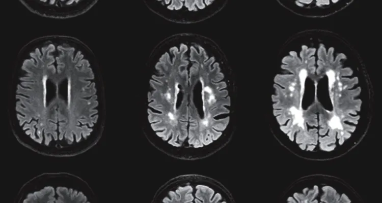
White matter hyperintensities are bright areas that appear on specific MRI sequences, particularly Fluid-Attenuated Inversion Recovery (FLAIR) imaging.
These bright spots on an MRI can signal blood vessel damage and ischemic injury. They are extremely common and become more prevalent with age, but WMH can arise from several causes, the most common of which is chronic small vessel disease, sometimes called chronic microvascular ischemic change.
WMH are not just incidental MRI findings. Research suggests these lesions represent areas of chronic vascular injury where blood flow has become impaired over time and injured the underlying brain, reinforcing their connection to long-term cardiovascular and brain health. [2]
Why do white matter hyperintensities matter?
Even though WMH findings are common as you age, that doesn't mean they don’t warrant intervention.
Research consistently demonstrates that greater WMH burden is associated with: [3] [4]
WMH are often the visible endpoint of cardiovascular risk factors that may have been developing silently for years. From a preventive medicine perspective, these bright lesions are actionable clues.
They show us that vascular injury is already present and that more aggressive management of underlying risk factors is likely needed to stave off further damage.
How does blood pressure affect the brain over time?
The number one cardiovascular risk factor most closely linked to white matter disease is blood pressure. [5]
The brain's small vessels are particularly vulnerable to chronic pressure-related injury. Over time, elevated blood pressure can damage vessel walls, impair blood flow regulation, and contribute to the development of ischemic white matter abnormalities.
But you may not even know this is happening to you, as early on it usually leads to little or no symptoms.
Many people tend to feel well while vascular damage gradually accumulates.
By the time cognitive symptoms appear, the opportunity for meaningful preventive measures is reduced. That’s one reason why blood pressure management remains among the most powerful interventions available for protecting both cardiovascular and cognitive health.
What is the connection between ApoB, LDL-C, and brain health?
Blood pressure isn’t the only factor involved in brain health. Your lipid profile plays a role, too.
Apolipoprotein-B (ApoB)-containing lipoproteins are central drivers of atherosclerosis throughout your vascular system. While ApoB and low-density lipoprotein cholesterol (LDL-C) are often discussed in the context of heart attacks and coronary artery disease, their effects extend beyond the heart.
Elevated ApoB doesn’t just increase heart disease risk. It also contributes to vascular injury throughout the body, including the vessels supplying the brain. [6]
What does "what's good for the heart is good for the brain" actually mean?
This phrase is commonly repeated because it is supported by decades of research. The same interventions that reduce cardiovascular risk also support cognitive longevity.
These include:
All of these interventions support your whole body. Specifically, they improve vascular health broadly, which ultimately benefits both heart and brain function.
How does Biograph combine cardiovascular risk assessments and neuroimaging in its Method?
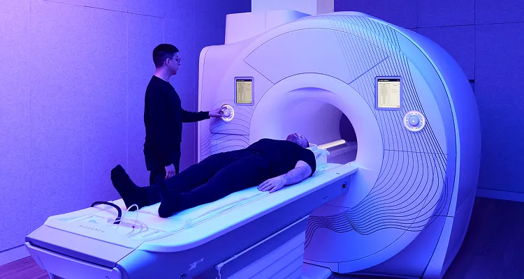
At Biograph, brain imaging is never interpreted in isolation. Whole-body MRI findings are evaluated alongside:
This integrated approach allows us to understand not only what appears on a scan, but why it may be occurring.
For example, a member with elevated WMH burden may also have an elevated ApoB level, elevated blood pressure, reduced cardiovascular fitness, and insulin resistance.
Viewed together, these findings provide a much clearer picture of long-term cardiovascular and cognitive risk than any single test alone, and identify areas for intervention.
Early detection only matters if it changes what happens next
Finding WMH is not the goal. Improving the trajectory of your future health is.
At Biograph, imaging findings are translated into actionable care plans focused on the upstream drivers of disease. Members work with physicians and specialists to improve cardiovascular fitness, optimize blood pressure, address lipid abnormalities, improve metabolic health, and monitor progress over time. This longitudinal approach is what transforms early detection into meaningful prevention.
The earlier we recognize those signals, the more opportunity we have to change health outcomes later down the line.
The bottom line
Brain MRI is often thought of as a neurological tool, but it’s also a powerful window into long-term cardiovascular health.
White matter hyperintensities provide visible evidence of cumulative vascular injury and can reveal brain injury long before cognitive symptoms appear.
The same factors that drive heart disease, such as high blood pressure, elevated ApoB, metabolic dysfunction, inflammation, and poor cardiovascular fitness, also influence cognitive aging and brain health.
At Biograph, advanced neuroimaging is combined with cardiovascular testing, metabolic assessment, and longitudinal follow-up to identify risk earlier and create targeted opportunities for intervention.
What are white spots on a brain MRI?
White spots are often called white matter hyperintensities (WMH). They commonly reflect small vessel disease and cumulative vascular stress affecting the brain's blood supply.
Do white matter hyperintensities mean dementia?
Not necessarily. WMH are associated with increased risk of cognitive decline, but many people with WMH never develop dementia. The findings should be interpreted within the broader context of cardiovascular and neurological health.
Can high blood pressure damage the brain?
Yes. Chronic high blood pressure is one of the strongest risk factors for white matter disease and can contribute to long-term vascular injury within the brain.
What does brain MRI reveal about cardiovascular health?
Brain MRI can reveal evidence of small vessel disease, prior silent strokes, and white matter changes that reflect the cumulative effects of cardiovascular risk factors over time.
Can brain imaging detect cardiovascular risk before symptoms appear?
In many cases, yes. A person may have no issues with memory or neurological symptoms but still show early signs of small vessel disease on imaging. Rather than waiting for those symptoms to emerge, clinicians can identify risk earlier and implement strategies designed to slow progression.
Can lifestyle changes improve vascular brain health?
Lifestyle changes cannot typically erase existing WMH, but improving blood pressure, cholesterol, metabolic health, fitness, nutrition, and sleep may help slow progression and reduce future risk.
Dr. Michael Doney is Biograph’s Executive Medical Director, with over 20 years of experience leading clinical care and advancing a more proactive, data-driven approach to medicine.
Clinical references
Brain Metabolism - an overview | ScienceDirect Topics. www.sciencedirect.com. https://www.sciencedirect.com/topics/medicine-and-dentistry/brain-metabolism
van Dalen JW, Mutsaerts HJMM, Nederveen AJ, Vrenken H, Steenwijk MD. White Matter Hyperintensity Volume and Cerebral Perfusion in Older Individuals with Hypertension Using Arterial Spin-Labeling. American Journal of Neuroradiology. Published 2016. Accessed June 23, 2026. https://www.ajnr.org/content/37/10/1824
Song R, Xu H, Dintica CS, et al. Associations Between Cardiovascular Risk, Structural Brain Changes, and Cognitive Decline. Journal of the American College of Cardiology. 2020;75(20):2525-2534. doi:10.1016/j.jacc.2020.03.053
Zhang S, Hu Y, Yang H, Li Q, Chen J, Bai H. Value of white matter hyperintensity volume and total white matter volume for evaluating cognitive impairment in patients with cerebral small-vessel disease. Frontiers in Aging Neuroscience. 2023;15. doi:10.3389/fnagi.2023.1096808
Fuhrmann D, Nesbitt D, Shafto M, et al. Strong and specific associations between cardiovascular risk factors and white matter micro- and macrostructure in healthy aging. Neurobiology of Aging. 2019;74:46-55. doi:10.1016/j.neurobiolaging.2018.10.005
Hu F, Wu R, Zhao X, et al. High Apolipoprotein B Levels are Associated with an Increased Risk of Recurrent Acute Ischemic Stroke: A Nested Case–Control Study. Translational Stroke Research. 2025;16(6):2035-2046. doi:10.1007/s12975-025-01367-9